Orbital Roof Fracture Radiology

Pin On Mr Radiology

Pin On Mr Radiology

Orbital Roof Blow In Fracture Radiology Case Radiopaedia Org

Orbital Roof Blow In Fracture Radiology Case Radiopaedia Org

Blowout Orbital Fracture Radiology Medical Imaging Radiography

Blowout Orbital Fracture Radiology Medical Imaging Radiography

Osteoma Of Orbital Roof Radiology Case Radiopaedia Org Radiology Brain Images Mri Brain

Osteoma Of Orbital Roof Radiology Case Radiopaedia Org Radiology Brain Images Mri Brain

Superior Orbital Fracture Radiology Case Radiopaedia Org

Superior Orbital Fracture Radiology Case Radiopaedia Org

Pathological Clavicle Fracture Radiology Case Radiopaedia Org Radiology Pathology Clavicle

Pathological Clavicle Fracture Radiology Case Radiopaedia Org Radiology Pathology Clavicle

Pathological Clavicle Fracture Radiology Case Radiopaedia Org Radiology Pathology Clavicle

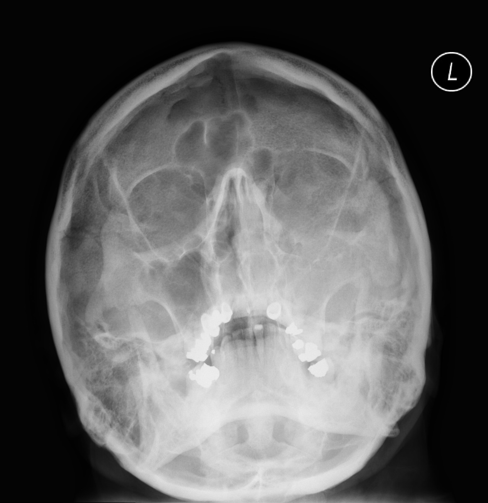
Communited mildly depressed left orbital roof fracture.

Orbital roof fracture radiology.

Orbital fracture pearls are as follows. Radiological considerations in the setting of orbital roof fracture it is important to fully assess the extent of the fracture including any radiographically apparent concomitant abnormalities. Displaced orbital roof fracture 3. Dr robert foley and assoc prof frank gaillard et al.

The orbital roof is composed of the orbital plate of the frontal bone with a small contribution from the lesser wing of the sphenoid at the apex figures 3 4 and 3 5. Fracture of the left zygomaticomaxillary attachmnet with fracture of the anterior and lateral maxillary walls inferior orbital rim fracture and left maxillary hemosinus. Angulated displaced fractures fragments are seen projecting downwards within the orbit indenting the superior rectus muscle. As an isolated orbital roof fracture is a rare entity it is important to.

Isolated orbital roof fractures in adults are uncommon comprising 12 19 of all orbital wall fractures. Fractures of the orbital roof are usually seen in combination with extension of linear frontal bone fractures or with complex cranial facial fractures including le fort iii naso orbito ethmoidal noe skull base fractures extending into the anterior skull base. Although common in the pediatric population isolated orbital roof fractures can also occasionally occur in an adult fig 13. It is a thin lamina separating the orbit anteriorly from the frontal sinus and posteriorly from the anterior cranial fossa.

Orbital blowout fractures occur when there is a fracture of one of the walls of orbit but the orbital rim remains intact. The injury can be associated with a violation of the dura necessitating an intracranial approach. When they do happen they may be associated with intracranial hemorrhages traumatic optic neuropathy and csf leakage into the orbit. This is typically caused by a direct blow to the central orbit from a fist or ball.

Osteoma Of Orbital Roof Radiology Case Radiopaedia Org Radiology Radiology Imaging Diagnostic Imaging

Osteoma Of Orbital Roof Radiology Case Radiopaedia Org Radiology Radiology Imaging Diagnostic Imaging

Superior Orbital Roof Blowout Fracture With Intact Orbital Rim Radiology Case Radiopaedia Org

Superior Orbital Roof Blowout Fracture With Intact Orbital Rim Radiology Case Radiopaedia Org

Radial Head Fracture Radiology Case Radiopaedia Org Radiology Fractures Medical School Stuff

Radial Head Fracture Radiology Case Radiopaedia Org Radiology Fractures Medical School Stuff

Pin On Radiology Spotters

Pin On Radiology Spotters

Facial Bone X Ray Lateral View Www Anatomynote Com Facial Bones Radiology Radiology Humor

Facial Bone X Ray Lateral View Www Anatomynote Com Facial Bones Radiology Radiology Humor

Fibrous Dysplasia Frontal A And Lateral B Plain Radiographs Show A Well Defined Thickened And Sclerotic Appearance Of The R Patient Radiology Radiographer

Fibrous Dysplasia Frontal A And Lateral B Plain Radiographs Show A Well Defined Thickened And Sclerotic Appearance Of The R Patient Radiology Radiographer

Multiple Myeloma Is The Most Common Primary Malignant Bone Neoplasm In Adults And Results In A Wide Range Of Ra Radiology Multiple Myeloma Pediatric Radiology

Multiple Myeloma Is The Most Common Primary Malignant Bone Neoplasm In Adults And Results In A Wide Range Of Ra Radiology Multiple Myeloma Pediatric Radiology

Derrick Rose Orbital Fracture Surgery Derrick Rose Plastic And Reconstructive Surgery Head And Neck

Derrick Rose Orbital Fracture Surgery Derrick Rose Plastic And Reconstructive Surgery Head And Neck

Sacrococcygeal Giant Cell Tumour Radiology Case Radiopaedia Org Tumor Radiology Cell

Sacrococcygeal Giant Cell Tumour Radiology Case Radiopaedia Org Tumor Radiology Cell

Blow Out Fracture The Most Common Portion Of The Orbit To Sustain A Fracture Is The Weak Floor And This Injury If Occurring In Isolation May Result In A Blow

Blow Out Fracture The Most Common Portion Of The Orbit To Sustain A Fracture Is The Weak Floor And This Injury If Occurring In Isolation May Result In A Blow

Maxillofacial Injuries Radiology Key

Maxillofacial Injuries Radiology Key

Pin On Radiologie

Pin On Radiologie

Pin En Radiology Interesting Cases And Spotters

Pin En Radiology Interesting Cases And Spotters

Vocal Cord Paralysis Radiology Reference Article Radiopaedia Org In 2020 Thyroid Surgery Paralysis Nerve Palsy

Vocal Cord Paralysis Radiology Reference Article Radiopaedia Org In 2020 Thyroid Surgery Paralysis Nerve Palsy

Chest Ct Scan In A Patient Who Was Born With Only One Lung Arrowhead Normal Lung Arrow Heart Shifted To Where O Diagnostic Imaging Radiology Ct Scan

Chest Ct Scan In A Patient Who Was Born With Only One Lung Arrowhead Normal Lung Arrow Heart Shifted To Where O Diagnostic Imaging Radiology Ct Scan

Colles Fracture Posterior Displacement

Colles Fracture Posterior Displacement

Burst Fracture Of Lumbar Spine Sagittal Reconstruction Of Ct Of The Lumbar Spine Demonstrates A Comminuted Vertical Burst Fracture Through The Body Of L1 Whit

Burst Fracture Of Lumbar Spine Sagittal Reconstruction Of Ct Of The Lumbar Spine Demonstrates A Comminuted Vertical Burst Fracture Through The Body Of L1 Whit

Mount Fuji Sign Is Seen On Cross Sectional Imaging And Implies Tension Pneumocephalus Is Present The Sign Refers To The Pre Radiology Sinusitis Brain Images

Mount Fuji Sign Is Seen On Cross Sectional Imaging And Implies Tension Pneumocephalus Is Present The Sign Refers To The Pre Radiology Sinusitis Brain Images

1

1

Pin On Surgery

Pin On Surgery

Sclerotic Bone Leison Note Sclerotic Bone Leison In Tibia In Young Age Osteoid Osteoma Note Malignancy In Old Age Radiology Tumor Oral Pathology

Sclerotic Bone Leison Note Sclerotic Bone Leison In Tibia In Young Age Osteoid Osteoma Note Malignancy In Old Age Radiology Tumor Oral Pathology

Subdural Acute On Chronic Radiology Case Radiopaedia Org Radiology Medical Imaging Brain Images

Subdural Acute On Chronic Radiology Case Radiopaedia Org Radiology Medical Imaging Brain Images

Infantile Head Trauma With Orbital Roof Fracture Radiology Case Radiopaedia Org

Infantile Head Trauma With Orbital Roof Fracture Radiology Case Radiopaedia Org

Pin On Radiology Study

Pin On Radiology Study

Genitourinary Radiology Kidney Mass Polycystic Kidneys Kidney

Genitourinary Radiology Kidney Mass Polycystic Kidneys Kidney

Https Radiopaedia Org Articles Bone Within A Bone Appearance 1 Radiology Pediatric Radiology Bone Diseases

Https Radiopaedia Org Articles Bone Within A Bone Appearance 1 Radiology Pediatric Radiology Bone Diseases

Granulomatosis With Polyangiitis Pulmonary Manifestations Radiology Reference Article Radiopaedia Org In 2020

Granulomatosis With Polyangiitis Pulmonary Manifestations Radiology Reference Article Radiopaedia Org In 2020

Full Size Picture 500178 Fx10 Jpg Artwork Full Size

Full Size Picture 500178 Fx10 Jpg Artwork Full Size

Is Case 332 Bronchial Atresia Radiology Bronchial Pet Ct

Is Case 332 Bronchial Atresia Radiology Bronchial Pet Ct

International Day Of Radiology Radiopaedia Org Radiology Progressive Supranuclear Palsy Radiology Imaging

International Day Of Radiology Radiopaedia Org Radiology Progressive Supranuclear Palsy Radiology Imaging

Pin On E

Pin On E

Plasmacytoma Radiology Case Radiopaedia Org Radiology Case Limb

Plasmacytoma Radiology Case Radiopaedia Org Radiology Case Limb

Peripheral Ossifying Fibroma Google Search Character Art Darth

Peripheral Ossifying Fibroma Google Search Character Art Darth

Pulmonary Sequestration Diagnostic Imaging Radiology Medical Knowledge

Pulmonary Sequestration Diagnostic Imaging Radiology Medical Knowledge

Pin On X Ray School Show

Pin On X Ray School Show

Pulmonary Aneurysm With Thrombus Inside Aneurysm Pulmonary

Pulmonary Aneurysm With Thrombus Inside Aneurysm Pulmonary

Pxa Or Extra Ventricular Supratentorial Ependymomoa Which Is Common To Show Cyst And Nodule Cysts Personalized Items Head And Neck

Pxa Or Extra Ventricular Supratentorial Ependymomoa Which Is Common To Show Cyst And Nodule Cysts Personalized Items Head And Neck

Adaugă Pin Pe Face And Neck

Adaugă Pin Pe Face And Neck

Gray Matter Heterotropia Cc Agenesis Radiology Head And Neck Gray Matters

Gray Matter Heterotropia Cc Agenesis Radiology Head And Neck Gray Matters

Neurofibromatosis Type Ii Radiology Case Radiopaedia Org Radiology Neurology Brain Images

Neurofibromatosis Type Ii Radiology Case Radiopaedia Org Radiology Neurology Brain Images

Orbital Blowout And Retro Orbital Hematoma Radiology Case Radiopaedia Org

Orbital Blowout And Retro Orbital Hematoma Radiology Case Radiopaedia Org

Pin By Eva On Mri In 2020

Pin By Eva On Mri In 2020

Pin By Mohamed Ouhlous On Neuro Dermoid Cyst Epidermoid Cyst Diagnostic Imaging

Pin By Mohamed Ouhlous On Neuro Dermoid Cyst Epidermoid Cyst Diagnostic Imaging

Https Encrypted Tbn0 Gstatic Com Images Q Tbn 3aand9gcqycbcdgoe6rzb6oic0fdozorujti4io1tvpnsexbak2qbljwr3 Usqp Cau

Https Encrypted Tbn0 Gstatic Com Images Q Tbn 3aand9gcqycbcdgoe6rzb6oic0fdozorujti4io1tvpnsexbak2qbljwr3 Usqp Cau

Source : pinterest.com